
Pattabiraman Lab
We study age-related ocular neuropathies including Glaucoma, the association of diabetes mellitus and ocular hypertension, and age-related macular degeneration.

Ocular Hypertension and Primary Open Angle Glaucoma Research
Research
The Pattabiraman laboratory studies the pathophysiology and treatment paradigms for primary open-angle glaucoma (POAG). Glaucoma is the second leading cause of blindness around the world and POAG is the leading form of glaucoma in the United States.
Glaucoma is manifested by the loss of peripheral vision. The clinical characteristic of glaucoma is an increased cup-to-disc ratio on retinal examination, which assesses the progression of glaucoma. This increased cup to disc ratio reflects the loss of retinal ganglion cell (RGC) axons due to the degeneration of RGCs. Ocular hypertension (OHT) or elevated intraocular pressure (IOP) is believed to be responsible for the pathogenesis of POAG and reducing IOP is the only means to prevent vision loss. OHT causes the RGC death due to the biomechanical stress imparted on the retina. This loss of vision is irreversible and lowering the IOP is the only mode of reversing the vision loss or halting the progression into blindness.
Under this program, we:
i) Investigate the cellular and molecular regulators of IOP homeostasis and identify pathological mechanisms leading to elevated IOP or OHT,
ii) Develop novel ocular therapeutics to lower OHT,
iii) Study the mechanistic evidence on how OHT modulates retinal ganglion cell (RGC) survival.
iv) Differences in the structure and function of trabecular outflow tissues between people of African and European Descent.
v) Understand the role of hyperglycemia and diabetes mellitus as a risk factor for OHT


News & Events

Heartland Vision Research Symposium
Best oral presenter award Srimathi Raghavan
August 2025


White Coat Ceremony for Idris Adekale
September 2025
SNRI Research Day
Best Presentation Winners
Roda Pasteurin and Anusha Shivanshankar
October 2025

Brightfocus-ISER Glaucoma FastTrack
Travel award winners -
Srimathi Raghavan & Anusha Shivashankar
October 2025

Yaay! Congratulation Roda on your F31.
Roda received her Notice of Award
July 7 2026

Way to go, Anusha, for being elected as the GRS Signaling by Adhesion Receptors 2028 Co-Chair.
June 2026

Avinash Soundararajan - First Big Grant from Knights Templar Eye Foundation - Career Starter Grant
April 2026

Adalyn DeWitt - Best Medicine Poster at the IUSM 2026 Spring Student Research Symposium
April 2026


Gregory Rognon, Postdoctoral Fellow from the lab matched at William Beaumont Ophthalmology Residency Program — many congratulations Greg.
Brightfocus-ISER Glaucoma FastTrack
Poster presenter award Srimathi Raghavan
October 2025


Postdoc Avinash and Graduate student Ting from the lab won the best poster awards under the postdoc and graduate student category in the Biochemistry Department Research Day in October 2022.


We will be the official tweeter at the #ASBMB2022


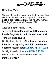
Our graduate student, Ting Wang's work on the role of cholesterol in regulating actin and cellular stiffness is accepted for an oral presentation at ASBMB 2022 in the SPOTLIGHT SESSION.
https://www.asbmb.org/meetings-events/2022-annual-meeting
Way to go Ting.
Latest Publications
